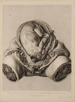
 As noted by Ludmilla Jordanova and Deanna Petherbridge in The Quick and the Dead: Artists and Anatomy, artists like Michelangelo and Leonardo da Vinci made enormous contributions to the emerging sciences of the body. The study of anatomy was, in fact, obligatory for many schools of art–and artists like Allessandro Allori composed anatomy textbooks for physicians.[i]
The close approximation of art and anatomy meant that the artists needed both “perceptual drawing skills” and “a strong stomach,”[ii] but just as the artist might be sometimes an anatomist, the anatomist or physician might sometimes be an artist. In this post, we will look at the work of Jan van Rymsdyk, the Dutch anatomy artist and his work with William Smellie and William Hunter.
Though the anatomical depiction of the 16th and 17th centuries tended to present whole bodies, often moving as though they were living, the figured in 18th century (particularly female anatomies) anatomical atlases are often shown piecemeal, as parts rather than whole. As Jordanova and Petherbridge point out, the rendering of sexual difference is very important in this period, especially as ideas about motherhood, breast feeding, and female responsibility were changing. As we have noted elsewhere on the blog, it is also the period when male practitioners took over midwifery. The man-midwife (usually also a surgeon) used tools—like the Chamberlen forceps—to aid in birth, and more and more complex anatomical images and models were produced to render the female anatomy as transparent as possible for the job.
One interesting consequence of this desire for clarity is the privileging of the infant body over the woman’s body. She often does not appear at all except by her fragments, a womb only. It is easy to read a narrative of displacement in these images: even as the midwife is replaced, so too is the mother. The machinery of the woman is laid bare (and of course, there are actually “woman machines” too—the devices built by Smellie to simulate a woman in labor).
“[Rymsdyk’s drawings] are amazingly powerful, not only for their subject matter but also in the confidence and beauty of their treatment […] The relationship in these images between the real and idealized, the whole and the fragment, and what is represented and what is repressed, therefore invited complex readings”[iii]
Rymsdyk often employed red chalk for his renderings, and with amazing detail and skill. By mixing dry chalk with wet chalk, stippling with strokes, he creates images so realistic that they seem strangely alive (or even ‘uncanny’ in their life-like familiarity). The original chalk drawings were part of Hunter’s library, and are presently housed at the University of Glasgow rare book collection. We probably know more about his techniques than his life history, though he was working in London by 1750 with William Hunter, and he was probably working simultaneously with Smellie. Rymsdyk later set out to be a portrait painter rather than a medical artist, but had minimal success.
Rymsdyk and Hunter
Though Jan van Rymsdyk’s first published work appears in William Smellie’s Sett [sic] of Anatomical Tables, published in 1754, he likely began work for Hunter in 1750. It would take more than 20 years to bring Hunter’s Gravid Uterus into print. The Hunterian Museum in Glasgow has a portfolio containing 61 drawings, including the provocative plate 6, wherein the womb is shown with amputated legs that appear, rather disturbingly, like two hams. A similar view is shown in Smellie’s tables (or plates), though in this image, Rymsdyk used the method of draping extremities with cloth. We might speculate whether this was artistic license or done at the behest of the physician, but plate 6 did earn Hunter’s praise.[iv]
Despite being a major contributor to the Gravid Uterus, Rymsdyk is not mentioned by name. That has led some to speculate about the rapport between the two men, but there is little evidence in print to document their working relationship.
Rymsdyk and Smellie
 We have only marginally more knowledge about Rymsdyk’s relationship with William Smellie, but there are certain clues that suggest Smellie respected Rymsdyk’s desire to be a portrait artist (often thought of as a more prestigious career). In 1753, Rymsdyk painted a portrait of Smellie. The original has subsequently been lost, but an engraving by Charles Grignion of the portrait remains (shown here). Interestingly, William Smellie also painted a portrait of himself—in his later years (below). He was an accomplished artist as well, and it is reasonably supposed that tables XXXVII and XXXIX of A sett of Anatomical Tables were the work of Smellie rather than Rymsdyk.[v]
Anything beyond this is mainly speculation. Perhaps, himself an artist, Smellie recognized the aspirations of  another. Smellie’s career was made as a surgeon and man-midwife; if he had once entertained notions of being a more accomplished or respected artist, these had long been left in the past. He leaves most of the illustration to Rymsdyk, and when he realized addition would be necessary, he engaged William Camper. We are left to speculate why Smellie decided to use his own work for the 37th and 39th plates, but both of these concern, specifically, the use of forceps, hooks, or crotchets as forceps. Given that Smellie was described by his pupils as “a mechanical genius,” and that he modified the forceps design, he perhaps felt best able to represent them.
Art and Practicality
The artistry of anatomy in the age before photography is complex and frequently suggestive. It may be part of the ongoing mission to move birth and reproduction firmly into the realm of science (and medical men). It may also be a reflection on changing understandings about the body–from a holistic model to one focused on specialization and the working of individual parts. Along the way, the artists and their works give us glimpses into the world of the medical theater (and occasionally, as with Jan van Rymsdyk’s desire to be a portrait painter, it tells the story of aspirations for something else or so
mething more). In any case, we can respect the incredible skill involved in creating the brilliant red-chalk images, so lifelike that they shame a camera lens. And yet, we must also return to the practical: though an artist drew the originals, the final publication included mainly engravings, the 18th-century version of the photo-copy (for a comparison, see Drawer of Wombs). In the words of Smellie himself: “The Whole of the Drawings were faithfully engraved (by Mr. Grignion); in which, however, delicacy and elegance have not been so much consulted as to have them done in a strong and distinct manner […] for general use.”[v]
[i] Jordanova, Ludmilla and Deanna Petherbridge. The Quick and the Dead: Artists and Anatomy. (1997): 8
[ii] Ibid., 14.
[iii] Ibid 6.
[iv] Thornton, John. Jan Van Rymsdyk. (1982): 32.
[v] Ibid., 15
About the blogger
Brandy Schillace is a medical humanist, literary scholar and writer of Gothic fiction. She is the Managing Editor for Culture, Medicine, and Psychiatry, a guest curator for Dittrick Museum, and a SAGES fellow for Case Western Reserve University (she has also worked as an assistant professor of literature at Winona State). She runs the Fiction Reboot and Daily Dose blogs, leads interdisciplinary conferences abroad for IDnet, and spends a lot of her time in museums and medical libraries.

